
“Vista Radiology is one of the first practices across the United States participating in ground-breaking medical trials of a new device used in large-diameter catheter procedures.”
– Dr. Jeffrey Roesch, Interventional Radiologist, Vista Radiology
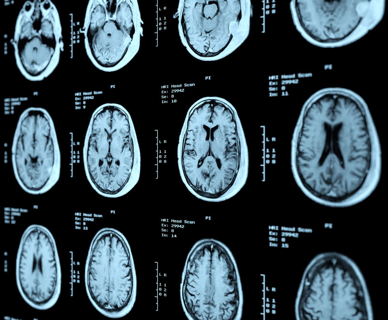
Computed Tomography (CT Scan, CAT Scan)
Computed Tomography (CT) – Abdomen and Pelvis
Computed Tomography (CT) – Angiography
Computed Tomography (CT) – Body
Computed Tomography (CT) – Chest
Computed Tomography (CT) – Head
Computed Tomography (CT) – Spine
Coronary Computed Tomography Angiography (CTA)
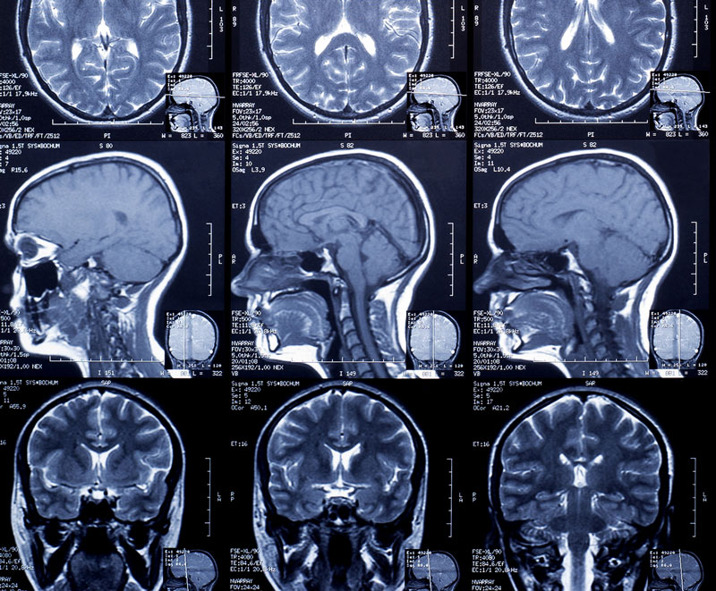
Magnetic Resonance Imaging (MR Scan, MRI Scan)
Magnetic Resonance (MR) – Angiography
Magnetic Resonance Imaging – Body
Magnetic Resonance Imaging – Cardiac (Heart)
Magnetic Resonance Imaging (MRI) – Chest
Magnetic Resonance Imaging (MRI) – Head
Magnetic Resonance Imaging (MRI) – Musculoskeletal
Magnetic Resonance Imaging (MRI) – Spine
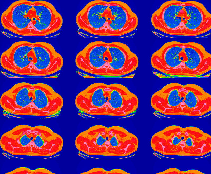
Nuclear Medicine and Positron Emission Tomography
(CT Scan, CAT Scan)
Nuclear Medicine, Cardiac
Nuclear Medicine, General
PET Scan
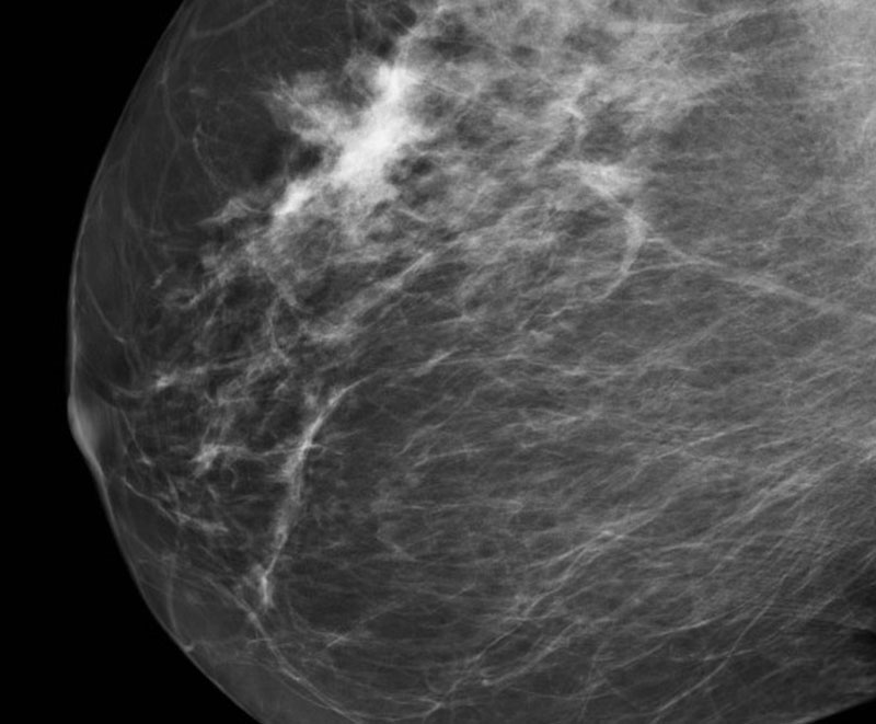
Women’s Imaging including Breast Imaging, Mammography, Breast MRI, Breast Biopsies and Bone Densitometry (DEXA)
Breast MRI
Breast Biopsy, MR-Guided
Breast Biopsy, Stereotactic
Breast Biopsy, Ultrasound-Guided
Breast Ultrasound
Mammography
Bone Densitometry
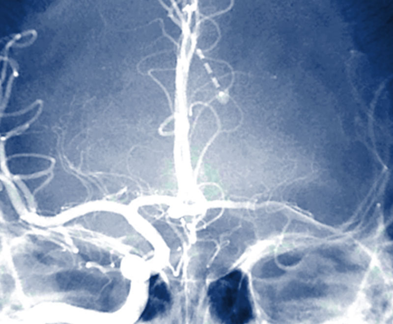
Interventional Radiology
Angioplasty and Vascular Stenting
Chemoembolization
Coil Embolization
Dialysis and Fistula/Graft
Declotting and Interventions
Embolization – Uterus (Fibroid Tumors)
Inferior Vena Cava Filter Placement and Removal
Myelogram
Nerve Blocks
Percutaneous Abscess Drainage
Radiofrequency Ablation of Kidney Tumors
Radiofrequency Ablation of Liver Tumors
Transjugular Intrahepatic Portosystemic Shunt (TIPS)
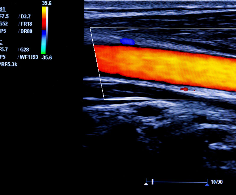
Ultrasound
Ultrasound – Abdomen
Ultrasound – Carotid
Ultrasound – General
Ultrasound – Musculoskeletal
Ultrasound – Obstetric
Ultrasound – Pelvis
Ultrasound – Vascular
Ultrasound – Venous
(Extremities)
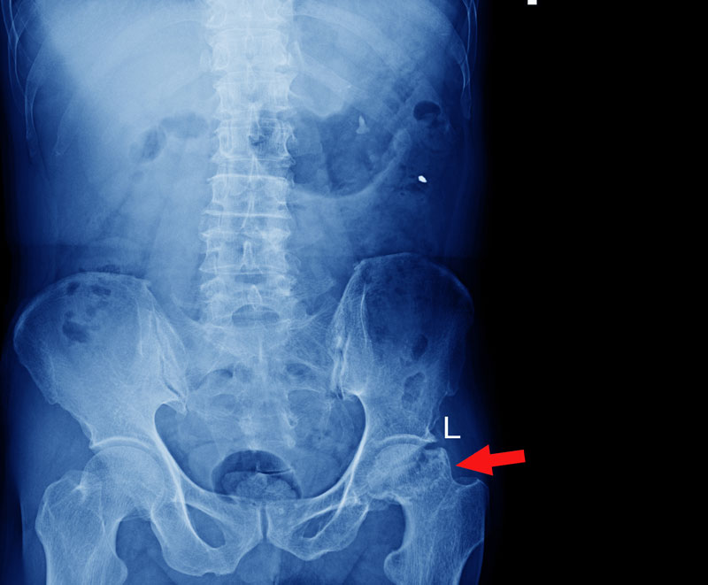
X-ray Imaging
Barium Enema
Barium Swallow
X-ray (Radiography), Bone
X-ray (Radiography), Chest
X-ray (Radiography), Lower GI Tract
X-ray (Radiography), Upper GI Tract
X-ray, Arthrography – Joints

